

SAMRI - Small Animal MRI Facility
High-resolution multimodal small animal imaging for preclinical research.
The SAMRI Core Facility provides state-of-the-art imaging technologies for mice and rats with a focus on ultra-high-field MRI and optical in vivo imaging. Researchers have access to cutting-edge infrastructure, scientific consultation, and comprehensive support for study design, data acquisition, and data analysis.
SAMRI supports preclinical research projects through advanced imaging technologies for small laboratory animals. The goal is to provide reproducible and high-quality data for translational research.
Core Technologies
- 11.7 Tesla small animal MRI
- IVIS Lumina systems for optical in vivo imaging
- Anatomical, functional, and molecular imaging
- Longitudinal studies in mouse and rat models
- Support for image analysis and data management
Scientific Focus
The facility supports projects in the following areas:
- Neuroscience
- Oncology
- Inflammation and immunology
- Metabolic diseases
- Pharmacological and translational research
Our Approach
SAMRI supports researchers from initial study planning to final data analysis:
- Consultation on the appropriate imaging method
- Support with animal models and experimental design
- Performance of imaging studies
- Data reconstruction and image analysis
Support for publications and grant applications
Equipment
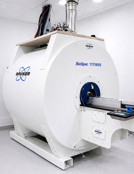
MRI System
- 11.7T preclinical Bruker MRI system
- Bruker BioSpec 117/16 platform
- BGS9 high-performance gradient system
- ParaVision 360 for acquisition, reconstruction, and system control
RF Coils & Specialized Hardware
Mouse Cryogenic Coil
- ¹H Quadrature Transmit/Receive Mouse Cryogenic Coil (CRP)
- Dedicated exclusively to mouse imaging
- Optimized for ultra-high-resolution imaging
- Particularly suitable for mouse brain imaging
- Significantly improved signal-to-noise ratio (SNR)
Volume Coils
- ¹H transmit-receive volume coils with active detuning
- Inner diameter: 72 mm
- ¹H transmit-receive volume coil
- Inner diameter: 40 mm
- Outer diameter: 75 mm
Surface Array Coils
- ¹H receive-only 2 × 2 mouse brain surface array coil
- ¹H receive-only 2 × 2 rat brain surface array coil
Physiological Monitoring
- Respiratory monitoring
- Cardiac monitoring
- Temperature control
Animal Handling Setup
- Isoflurane anesthesia system
- Animal beds with fixation and integrated heating
MRI Applications
- Structural MRI
- Functional MRI (fMRI)
- Diffusion MRI (DWI/DTI)
- Angiography
- MR spectroscopy
- Ex vivo imaging
- Longitudinal imaging studies
Additional Imaging Systems
- Two IVIS Lumina II systems for quantitative fluorescent and bioluminescent imaging
- Suitable for non-invasive longitudinal imaging in small animals
- Supports in vivo and ex vivo optical imaging applications
Software & Data Analysis
- ParaVision 360 for acquisition, reconstruction, system control, and image analysis
- Image post-processing software
- Quantitative data analysis tools
- Centralized data storage and reconstruction servers
- Analysis workstations
Services
Scientific Consultation
- Selection of suitable imaging methods
- Support with study design
- Advice on animal models and contrast agents
- Support with TVA-related aspects of animal approvals
Imaging Services
- Full-service imaging performed by facility staff
- Assisted self-use after training
- Standardized protocols
- Individually tailored imaging sequences
- In vivo and ex vivo imaging
- MR spectroscopy studies
Data Analysis
- Image reconstruction
- Segmentation
- Quantitative analysis
- Statistical analysis
- Support with figures and publications
Training
- Equipment introduction
- Safety training
- Introduction to image analysis software
- Individual workshops
Access and Booking
The facility is open to internal and external researchers from academic institutions, clinics, and industry partners.
Workflow
- Initial consultation and project discussion
- Selection of suitable imaging methods
- Scheduling
- Performance of imaging studies
- Data delivery and analysis
Requirements
- Approved TVA (animal experiment approval)
- Completed safety training
- Signed user agreement
- Compliance with SOPs
Booking
Appointments are coordinated through the central booking system of the institution.
Terms of Use and Pricing
Terms of Use
- Systems must be booked in advance.
- Users may only operate systems after training by SAMRI staff.
- MRI and IVIS systems may only be used according to approved procedures.
- Animals, samples, and potentially hazardous materials must be discussed with SAMRI staff before experiments.
- Users are responsible for compliance with TVA approvals, biosafety regulations, and animal welfare regulations.
- Data, samples, and consumables must be removed after each session.
- Independent use is possible only after successful training and approval by SAMRI staff.
Pricing
SAMRI operates on a cost-recovery basis.
The final cost depends on:
- MRI or IVIS instrument time
- Type of imaging application
- In vivo versus ex vivo experiments
- Level of staff support required
- Training sessions
- Data reconstruction and analysis
- Internal versus external users
Please contact the SAMRI staff for detailed pricing information and an individual quotation.
Downloads
Available Documents
- Terms of Use E | D
- SOPs (upon request)
- Safety documents
FAQ
Which animal species can be imaged?
Typically mice and rats. Other species may be possible upon request.
Can external users access the facility?
Yes, external academic and industrial partners are welcome.
Is support available for TVA applications?
Yes, the team supports imaging-related aspects of TVA applications and animal experiment approvals.
Can I operate the equipment independently?
Assisted self-use is possible after successful training.
Which data formats are provided?
Depending on the system, formats may include DICOM, NIfTI, TIFF, and raw data formats.
Contact
Contact Information
SAMRI – Small Animal MRI Core Facility
Head of SAMRI - Dr. Cornelia Hesse
Assistant - Carmen Rudert