
Detailed Insights with the World’s Most Powerful Brain Scanner
DZNE Expertise Delivers “Sharp” Images
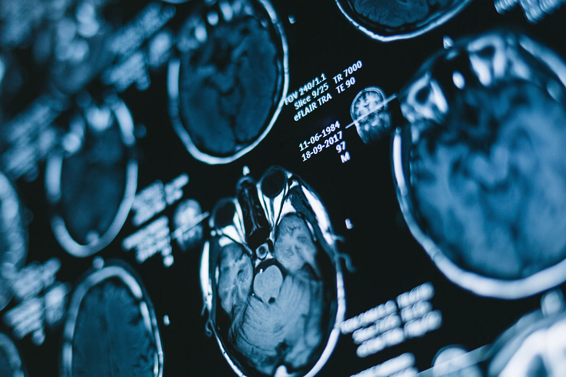
Bonn (Germany)/Paris (France), September 22, 2026. DZNE Researchers and French colleagues have presented high-resolution images of the human brain at a magnetic field strength of 11.7 Tesla. With a resolution of 300 micrometers (0.3 millimeters) acquired in less than 6 minutes, these are the most detailed images of the whole brain, including the basal ganglia to date. These neurological structures are located deep within the head. Potential applications of the applied technology include the diagnosis and treatment of Parkinson’s disease. The corresponding report was published in the scientific journal JAMA Neurology.
The images were acquired using the world’s strongest human MRI scanner, located at the research center “NeuroSpin” near Paris. The study utilized measurement protocols developed by DZNE. “Our pulse sequences enable high-resolution imaging in a short time, which is important for patient comfort. At the same time, motion artifacts - in other words: image distortions caused by head movement - are minimized by a novel technology implemented and refined in advance by my colleague Matthias Serger. The higher the resolution, the more critical this becomes,” explains DZNE researcher Dr. Rüdiger Stirnberg.
The brain structures captured in these images - the basal ganglia - are damaged in Parkinson’s disease. “The technology is still in the experimental stage. But in the long term, such high-resolution brain scans could be very useful for the diagnosis and treatment of Parkinson’s,” says Stirnberg.
Original publication
Unprecedented In Vivo MRI Resolution of Basal Ganglia at 11.7 T.
Béchir Jarraya et al.
JAMA Neurology (2026).
DOI/URL: https://doi.org/10.1001/jamaneurol.2026.3264