A sharper look into the head
An upgrade of magnetic resonance imaging (MRI) technology allows ten times more precise insights into the brain than before. Researchers involving DZNE report on this in the journal Nature Methods.
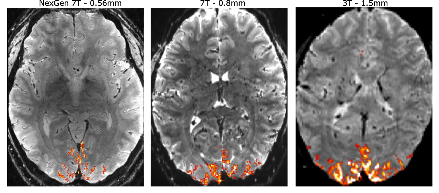
MRI has had a solid place in medical care and science for years – this applies in particular to brain research. This is because this technology provides detailed insights into the human body by leveraging magnetic fields and pulsed radio waves. MRI scanners that operate with a magnetic field strength of three Tesla are considered the gold standard in routine operation and scanners with a field strength of seven Tesla as state-pf-the-art, as they currently achieve the best spatial resolution. This applies particularly for so-called “functional MRI”, which localizes brain activity. An international team including Dr. Rüdiger Stirnberg, scientist at DZNE’s Bonn site, has now boosted this precision tenfold, making it possible to capture activity within brain areas measuring just around 0.4 millimetres. The researchers used an MRI device operated at the University of California in Berkeley, which they transformed into a “next-generation 7 Tesla scanner”. In addition to research institutions from the USA, Europe and South Korea, several companies were also involved in the multi-year project.
New hardware
“This is almost entirely a new development. Although the main field of seven Tesla and the associated magnetic coil have essentially remained the same, other hardware components have been completely redesigned. This applies in particular to the so-called gradient coils and the receiver system with which the signals from the human body are recorded. In a way, this is a very special radio receiver,” says Stirnberg.
Coordinated pulses
Important parts of the software have also been newly engineered. Specifically, this refers to the control of the radio pulses and additional magnetic fields – called “magnetic gradient fields” – that are required for image generation. “This can all be controlled by software. During an examination, this program runs automatically and switches gradient fields and radio pulses in the predefined manner and succession. This is called an MRI sequence,” says the Bonn physicist. “Colleagues from the USA and the Netherlands have used our sequences developed and adapted in Bonn in order to get the most out of the new technology. The various parameters have to be precisely coordinated. There is a lot of leeway.”
Smaller than a pinhead
Functional magnetic resonance imaging – fMRI for short – benefits particularly from the new developments. “With traditional fMRI, brain activity can be resolved spatially by recording signal changes due to local oxygen consumption. This is a marker of activity, because neurons need oxygen,” says Stirnberg. Novel fMRI methods, that rely on corresponding changes in blood volume, are locally even more precise. “To his end, many scientists worldwide use our sequence. In Berkely, this is now possible with unprecedented resolution. It’s like having focused a microscope. We can now measure the activity within tiny brain volumes that are only around 0.1 microliters in size. Such brain sections are smaller than a pinhead and typically contain less than a thousand neurons.”
Helpful for dementia research
Brain research in general and dementia research in particular should benefit from the new capabilities. There is also a transfer of experience and technology: Although the “NexGen 7 Tesla Scanner” in Berkeley is unique, the findings from this initiative will benefit both commercial development and the operation of existing devices. Stirnberg sees the project as a win-win situation for DZNE: “In Bonn, we operate a 7 Tesla scanner ourselves. We have gained valuable experience for our own studies from the development of the MRI sequences. Also, a novel scanner, in parts very similar to that in Berkeley, has recently been installed in Magdeburg. The scientists in Magdeburg are interested in our sequences and we are hoping to collaborate on advancing and using them there in the future.”
Original publication
Next-generation MRI scanner designed for ultra-high-resolution human brain imaging at 7 Tesla.
David A. Feinberg et al.
Nature Methods (2023)
DOI: 10.1038/s41592-023-02068-7
December 2023