New method for cell investigation
A unique technique enables researchers in Tübingen to follow what happens in brain cells in neurodegenerative diseases ̶ as if they were watching a live broadcast: In a research project that is now starting, the scientists will be able to age human brain tissue in a targeted manner in the Petri-dish.
Using a unique investigation method, researchers from Tübingen want to find out which role the so-called glial cells in the brain play in the development of neurodegenerative diseases. To achieve this, they are working with human tissue that had to be removed during head surgery. This innovative approach is funded by the U.S.-based Chan Zuckerberg Initiative with 1.6 million US dollars. The Hertie Institute for Clinical Brain Research, the University of Tübingen, the Department of Neurology at the University Hospital RWTH Aachen and the DZNE are involved in the study.
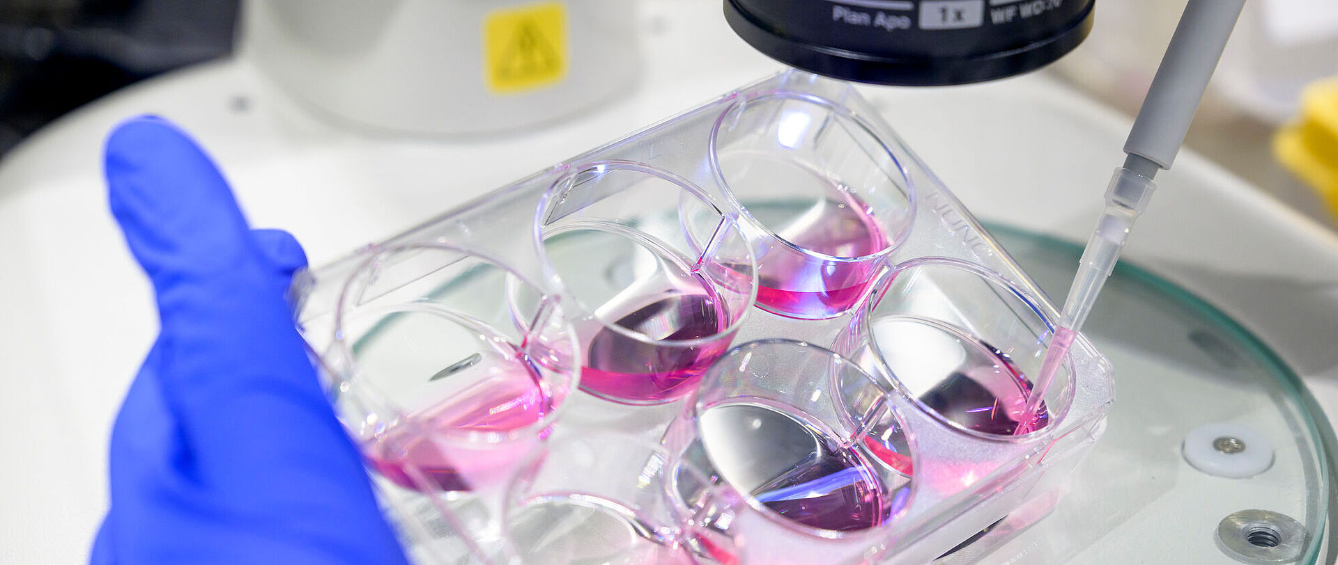
The focus of the studies is on wafer-thin sections of human tissue that are only 300 micrometers thick. "This is healthy tissue, but it blocks the way for physicians when they perform surgery on a brain tumor or an epileptic focus. Therefore, it has to be removed," said study lead Dr. Deborah Kronenberg-Versteeg, who conducts research at the DZNE site in Tübingen, within the research group around Prof. Jucker, the Hertie Institute for Clinical Brain Research and the University of Tübingen. The special feature: in the incubator for tissue cultures, the cellular processes in the tissue continue for weeks, even long after removal from the body. This is precisely the time the researchers are using, if the patients agree.
Among brain researchers, the hypothesis that neuronal dysfunctions are primarily caused by changes in glial cells has gained ground in recent years. Glial cells are a type of auxiliary cells that are arranged around neurons. They supply neurons with nutrients and all the other substances they need to do their work. If these glial cells no longer work properly, the neurons suffer ̶ and they are responsible for processing information in the brain. The scientists are now investigating this causal chain from the auxiliary cells to the neurons.
To do this, they are investigating what happens when they specifically cause the cells to become diseased. They drip a liquid containing so-called seeds onto the cultured tissue. These are small clumps of misfolded proteins. They cause pathological changes in neurons, leading to neurodegenerative diseases such as Alzheimer's or Parkinson's disease. "In the experiment, we add these misfolded proteins artificially; but in patients, they arise directly in the body," explained Deborah Kronenberg-Versteeg. "These seeds spread over the entire tissue in a very short time, like a snowball effect."
This is precisely the moment when the most exciting phase begins for her and her team: "We can observe on a live basis how the tissue reacts to this impact. Do deposits form? How quickly does that happen? And also: is young tissue more capable to break down the misfolded proteins than old tissue?" The fact that they work with tissue sections from real patients brings both advantages and disadvantages: Among the disadvantages is that the findings can only be generalized to a limited extent because the tissue is highly individual. On the other hand, this is precisely why samples from people of different ages are available. For example, it is possible to investigate whether the cells of 30-year-olds react differently from those of 80-year-olds - in other words, whether the brain's ability to respond well to a pathology declines with age.
"Worldwide, we are the only scientists who can look directly at these processes in human tissue, thanks to our newly developed method," Kronenberg-Versteeg said. The researchers involved had developed the method with the brain tissue cultures themselves. To do so, they had already received initial financial support from the Chan Zuckerberg Initiative, which was launched by Facebook founder Mark Zuckerberg and his wife Dr. Priscilla Chan. Funding was provided to 30 teams of scientists worldwide to develop innovative ideas in the field of neurodegenerative diseases. Subsequently, 16 of them were selected for the next funding period - among them the group from Germany, which, in addition to Deborah Kronenberg-Versteeg, also includes the research groups of Dr. Thomas Wuttke from the Hertie Institute for Clinical Brain Research and the University of Tübingen, and Dr. Henner Koch from the University Hospital RWTH Aachen. In the coming months, the team plans to use the new method to examine tissue sections from around 120 patients.